
Procedures
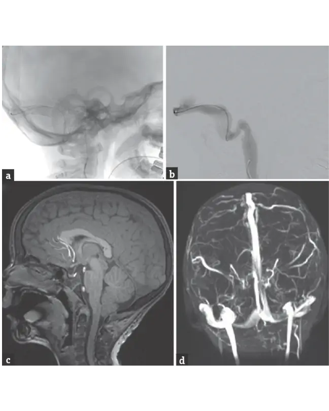
1. Venous Sinus Stenting (Refractory Intracranial Hypertension)
Procedure Overview
A stent is placed in narrowed venous sinuses of the brain to improve blood outflow and reduce intracranial pressure.
Procedure Preparation
- MRI/MRV and venography
- Pressure gradient measurement
- Blood tests and ophthalmic evaluation
- Consent
Pre-Treatment Procedure
- Assessment of vision and symptoms
- Confirmation of venous sinus stenosis
- Stabilization of intracranial pressure
Post-Treatment Care
- Monitoring neurological and visual status
- Antiplatelet therapy
- Follow-up imaging
Procedure Benefits
- Relieves headache and vision problems
- Prevents vision loss
- Minimally invasive
- Improves quality of life
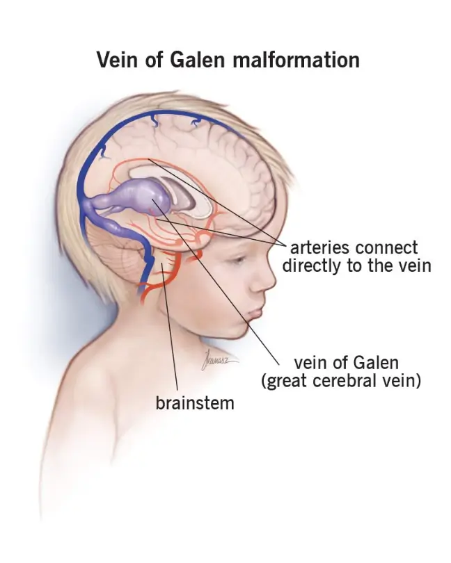
2. Vein of Galen Malformation (VOGM) Embolisation
Procedure Overview
Endovascular embolisation to block abnormal blood vessels in vein of Galen malformation, usually in infants or children.
Procedure Preparation
- MRI/CT and angiography
- Cardiac and neurological assessment
- Blood work
- Consent from guardians
Pre-Treatment Procedure
- Evaluation of heart failure and neurological status
- Planning staged embolisation
- Stabilization in ICU if needed
Post-Treatment Care
- NICU/ICU monitoring
- Developmental follow-up
- Repeat embolisation if required
Procedure Benefits
- Life-saving in critical cases
- Reduces abnormal blood flow
- Improves neurological outcomes
- Avoids open surgery
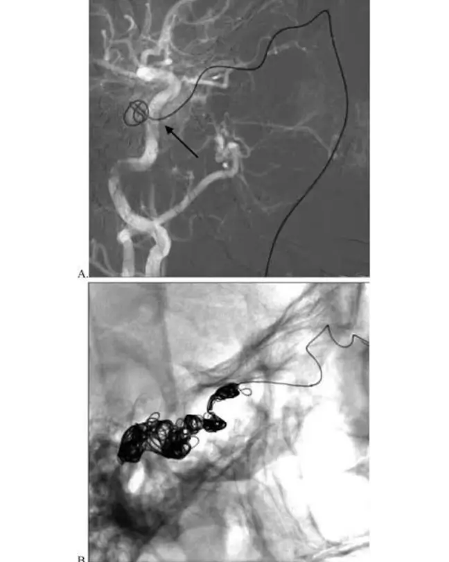
3. Carotid Cavernous Fistula (CCF) Coiling & Embolisation
Procedure Overview
A minimally invasive technique using coils or embolic agents to close abnormal connections between carotid artery and cavernous sinus.
Procedure Preparation
- CT/MRI and angiography
- Eye examination
- Blood tests
- Consent
Pre-Treatment Procedure
- Assessment of vision and eye symptoms
- Localization of fistula
- Hemodynamic
- stabilization
Post-Treatment Care
- Monitoring vision and neurological status
- Follow-up imaging
- Symptom resolution tracking
Procedure Benefits
- Relieves eye redness, swelling, and vision issues
- Prevents complications
- Minimally invasive with high success rate
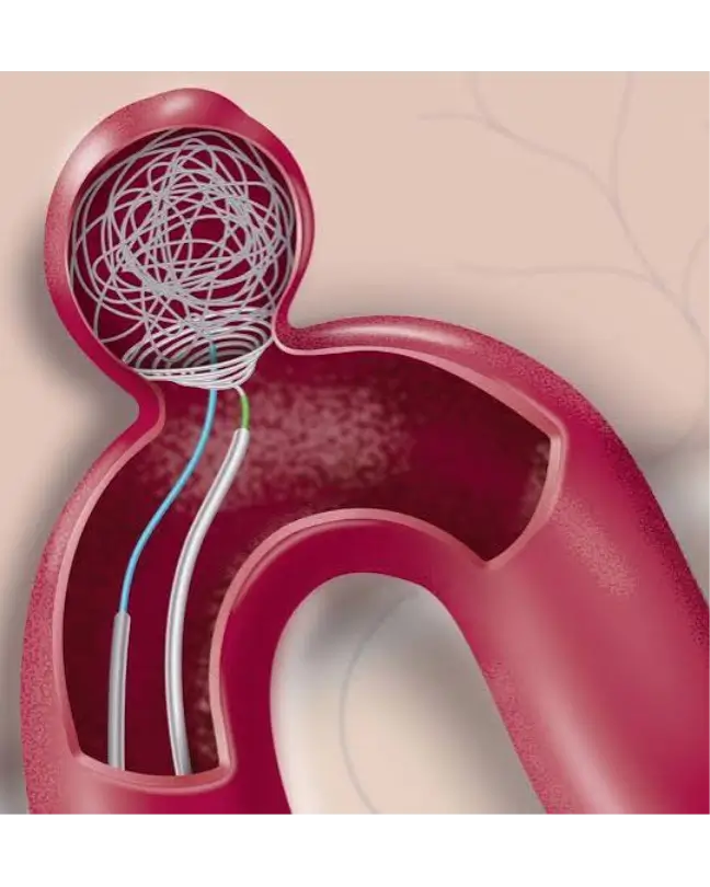
4. Intrasaccular Devices for Brain Aneurysms (WEB, Contour)
Procedure Overview
Devices like WEB or Contour are placed داخل aneurysm sac to block blood flow and prevent rupture.
Procedure Preparation
- CT angiography / DSA
- Blood investigations
- Assessment of aneurysm size and location
- Consent
Pre-Treatment Procedure
- Risk assessment for rupture
- Planning device size and approach
- Stabilization of patient
Post-Treatment Care
- Neurological monitoring
- Follow-up angiography
- Medication if required
Procedure Benefits
- Prevents aneurysm rupture
- Minimally invasive
- Reduced need for long-term medication
- Short recovery time
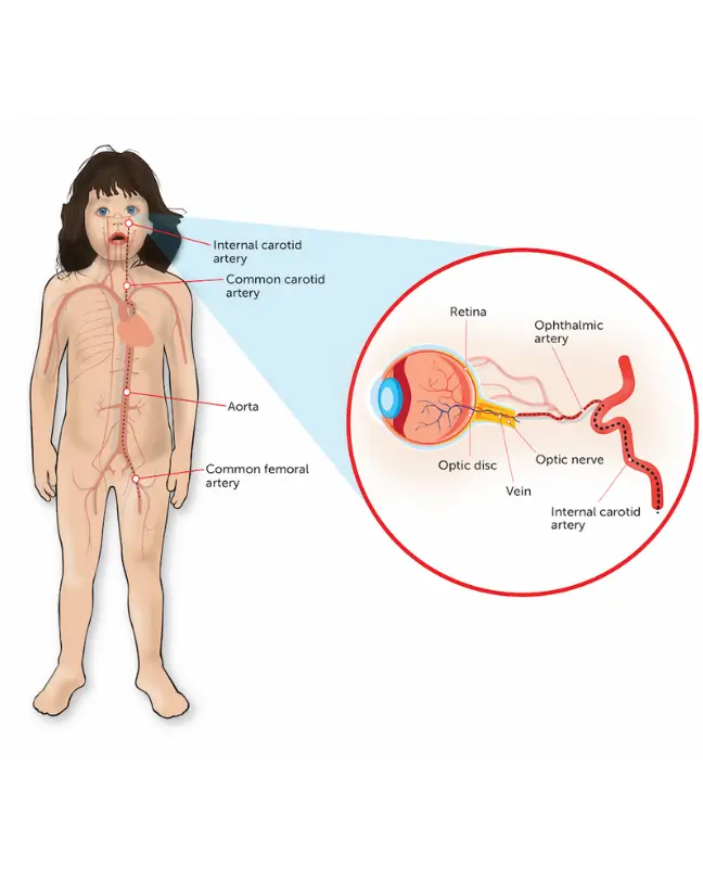
5. Intra-Arterial Chemotherapy for Retinoblastoma
Procedure Overview
Chemotherapy is delivered directly into the ophthalmic artery to treat eye cancer (retinoblastoma).
Procedure Preparation
- Eye examination under anesthesia
- Imaging studies
Blood tests - Consent from guardians
Pre-Treatment Procedure
- Tumor staging
- Planning drug dosage
- Patient stabilization
Post-Treatment Care
- Eye monitoring
- Repeat treatment cycles
- Vision assessment
Procedure Benefits
- Targeted therapy with fewer side effects
- Eye and vision preservation
- Reduced systemic toxicity
6. Preoperative Embolisation (Brain, Neck, Spine Tumors)
Procedure Overview
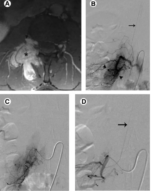
Tumor blood supply is blocked before surgery to reduce bleeding during tumor removal.
Procedure Preparation
MRI/CT and angiography
Blood work
Surgical planning
Consent
Pre-Treatment Procedure
Identification of tumor feeders
Hemodynamic assessment
Stabilization
Post-Treatment Care
Monitoring before surgery
Proceed to planned tumor surgery
Imaging if needed
Procedure Benefits
Reduces surgical blood loss
Improves surgical safety
Shortens operative time
7. Percutaneous Vertebroplasty / Kyphoplasty / Spinal Biopsy
Procedure Overview
Minimally invasive procedures to stabilize vertebral fractures or obtain tissue samples using needle-based techniques.
Procedure Preparation
Spine imaging (MRI/CT/X-ray)
Blood tests
Pain assessment
Consent
Pre-Treatment Procedure
Localization of affected vertebra
Assessment of fracture or lesion
Patient positioning
Post-Treatment Care
Pain monitoring
Early mobilization
Follow-up imaging
Procedure Benefits
Rapid pain relief
Spine stabilization
Minimally invasive
Quick recovery

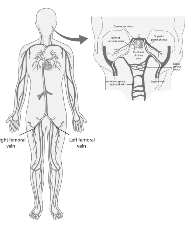
8. Inferior Petrosal Sinus Sampling (IPSS)
Procedure Overview
A diagnostic procedure to measure hormone levels from veins draining the pituitary gland to identify source of Cushing’s syndrome.
Procedure Preparation
Hormonal evaluation
Blood tests
Imaging of pituitary
Consent
Pre-Treatment Procedure
Catheter placement planning
Baseline hormone sampling
Patient stabilization
Post-Treatment Care
Monitoring for complications
Hormonal result interpretation
Endocrinology follow-up
Procedure Benefits
Accurate diagnosis of hormone source
Guides treatment planning
Minimally invasive diagnostic tool