
Digital Subtraction Angiography (DSA)
Advanced Cerebral & Spinal Vascular Imaging
Digital Subtraction Angiography (DSA) is a minimally invasive imaging procedure that provides highly detailed images of blood vessels in the brain, head, neck, spine, and spinal cord.
Using a thin catheter, contrast material, and real-time X-ray imaging, DSA helps specialists identify abnormal blood vessels, understand blood-flow patterns, and plan precise neurovascular treatment.
- High-Resolution Vascular Imaging
- Cerebral & Spinal Angiography
- Precise Treatment Planning
What Is DSA?
Digital Subtraction Angiography (DSA) is an advanced X-ray-based angiographic technique used to visualize blood vessels with exceptional clarity.
During the procedure, a small amount of contrast material is introduced into the blood vessels through a thin catheter. The imaging system captures detailed images as the contrast moves through the arteries and veins.
The “subtraction” technique digitally removes structures such as bone from the images, allowing the blood vessels to be seen more clearly.
Why DSA Is Important
DSA can provide detailed information about:
- The exact location of abnormal blood vessels
- The size and shape of vascular abnormalities
- The direction and pattern of blood flow
- Connections between arteries and veins
- Blockages, narrowing, or abnormal vessel anatomy
This detailed vascular information can help specialists determine the most appropriate treatment strategy.
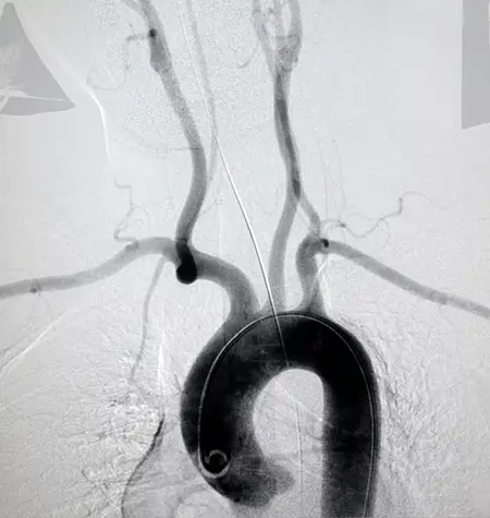
Cerebral DSA
Cerebral DSA examines the blood vessels supplying the brain, head, and neck.
It can help evaluate conditions such as:
- Brain aneurysms
- Brain AVMs and AVFs
- Blood vessel narrowing or blockage
- Vascular dissection
- Certain causes of stroke
- Abnormal blood-vessel patterns
- Blood supply to selected brain or head-and-neck tumors
Cerebral angiography can also provide detailed vascular mapping before or during certain neurovascular treatments.
Spinal DSA
Spinal DSA examines the arteries and veins surrounding the spinal cord and spine.
It is particularly useful for identifying and mapping:
- Spinal arteriovenous malformations (AVMs)
- Spinal dural arteriovenous fistulas (dAVFs)
- Abnormal spinal blood vessels
- Certain vascular causes of spinal cord problems
- Blood-vessel involvement around spinal or spinal-cord tumors
Because many spinal vascular abnormalities involve very small and complex vessels, detailed angiographic imaging can be important for accurate diagnosis and treatment planning.

When Is DSA Recommended?
Detailed Imaging for Complex Vascular Conditions
DSA may be recommended when non-invasive imaging such as CT angiography (CTA), MR angiography (MRA), CT, or MRI does not provide enough information, or when precise vascular mapping is required before treatment.
It may be used to:
- Confirm or further evaluate a suspected vascular abnormality
- Map an AVM or AVF before embolization
- Evaluate an intracranial aneurysm
- Assess complex blood-vessel narrowing or blockage
- Investigate certain unexplained neurological or vascular findings
- Plan minimally invasive neurovascular treatment
- Evaluate spinal vascular malformations
In selected cases, diagnostic angiography and an endovascular treatment may be performed during the same overall procedure when clinically appropriate.
How Is DSA Performed?
A Minimally Invasive Catheter-Based Procedure
DSA is performed in a specialized angiography suite under continuous imaging guidance.
Step 1 — Vascular Access
After local anesthesia, a thin catheter is introduced through a blood vessel, commonly through the wrist or groin, depending on the procedure and clinical situation.
Step 2 — Catheter Navigation
The catheter is carefully guided through the blood vessels to the area being examined.
Step 3 — Contrast Injection
A contrast material is injected through the catheter. As it travels through the blood vessels, high-resolution X-ray images are captured in real time.
Step 4 — Detailed Vascular Mapping
The images show the movement of blood through the arteries and veins, helping the specialist identify abnormal connections, narrowing, blockages, aneurysms, or other vascular abnormalities.
Step 5 — Recovery & Observation
After the catheter is removed, the access site is closed and the patient is monitored for a period before discharge or further care, depending on the procedure and individual condition.
Benefits, Safety & Treatment Planning
Precision That Supports Better Decisions
DSA provides highly detailed vascular images that can be particularly valuable when treatment decisions depend on understanding the exact anatomy and blood-flow pattern of a vascular abnormality.
Key Benefits
- Detailed visualization of brain and spinal blood vessels
- Precise mapping of complex vascular abnormalities
- Real-time assessment of blood flow
- Helps guide minimally invasive treatment
- Can provide information that complements CT or MRI-based angiography
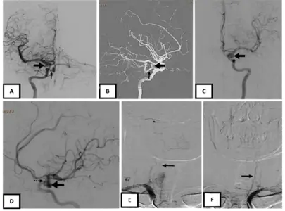
Safety & Considerations
DSA is generally well tolerated, but because it involves catheter access, contrast material, and X-rays, there are potential risks. These can include bruising or bleeding at the access site, infection, contrast reactions, blood-vessel injury, kidney-related complications in susceptible patients, and rarely stroke or other serious complications. Your specialist will assess your medical history, kidney function, medications, allergies, and other relevant factors before the procedure.
Advanced Imaging. Precise Understanding. Targeted Treatment.
Cerebral and spinal DSA provides the detailed vascular information specialists need to diagnose complex neurovascular conditions and plan treatment with greater precision.